
At ADR Spine, MRI imaging is a foundational tool in understanding spinal health and guiding treatment decisions. Led by founder Dr. Todd Lanman, a board-certified spinal neurosurgeon with decades of experience in motion-preserving spine surgery, the team uses advanced imaging to move beyond symptoms and identify the true structural cause of pain.
In this article, you will learn what a spine MRI is, how it works, which conditions it can diagnose, how it compares to other imaging studies, and when it may be time to consider getting one.
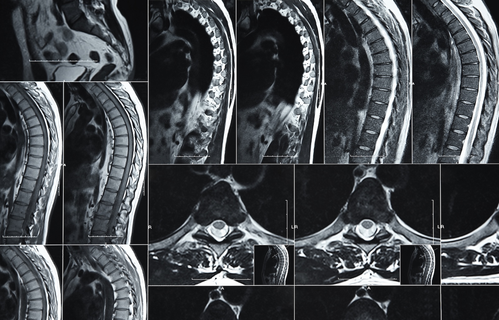
What Is a Spine MRI and How Does It Work?
An MRI is a non-invasive imaging test that uses powerful magnets and radio waves to create highly detailed, three-dimensional images of the spine. Unlike X-rays or CT scans, MRI does not use ionizing radiation.
This technology enables physicians to visualize soft tissues with exceptional clarity, including spinal discs, nerves, ligaments, muscles, and the spinal cord. Because many spinal conditions involve nerve compression or disc degeneration, MRI is often the most informative diagnostic tool available.
Common Spinal Conditions Diagnosed via MRI
Herniated or Bulging Discs
MRI is particularly effective at identifying disc problems. It can clearly show when the soft inner portion of a disc protrudes outward and presses on nearby nerve roots. This type of compression often explains symptoms such as radiating arm or leg pain, numbness, or weakness.
Spinal Stenosis
Spinal stenosis refers to the narrowing of the spinal canal or nerve passageways. On MRI, this appears as crowding around the spinal cord or nerve roots. MRI allows physicians to assess both the severity and the precise location of narrowing, which is critical for treatment planning.
Degenerative Disc Disease (DDD)
MRI scans reveal disc dehydration, height loss, and structural changes associated with degeneration. These findings help distinguish age-related changes from degeneration that is contributing to pain or nerve compression.
Spinal Tumors and Infections
MRI is the most sensitive imaging modality for detecting spinal cord tumors, metastatic disease, and spinal infections such as osteomyelitis or discitis. It allows early identification of abnormalities that may not be visible on other imaging studies.
MRI vs. X-Ray vs. CT Scan: Which Do You Need?
When X-Rays Are Sufficient
X-rays are often used as an initial imaging tool. They are effective for evaluating bone alignment, fractures, scoliosis, and instability. However, X-rays do not show discs, nerves, or soft tissues.
Why Your Doctor Might Order a CT Scan
CT scans provide detailed images of bone and are useful for complex fractures or surgical planning. They may also be used for patients who cannot undergo an MRI due to certain implanted devices. CT scans involve radiation and are less effective for evaluating nerves and discs.
The Superiority of MRI for Soft Tissue
MRI remains the gold standard for visualizing nerve roots, spinal discs, ligaments, and the spinal cord. It is often the preferred test when symptoms suggest nerve compression or when prior treatments have not resolved pain.
When Is It Time to Get a Spine MRI?
A spine MRI may be recommended if you experience:
- Persistent neck or back pain lasting more than several weeks
- Pain that radiates into the arms or legs
- Numbness, tingling, or muscle weakness
- Difficulty with balance or coordination
- Worsening symptoms despite conservative treatment
Urgent imaging may be necessary if symptoms include loss of bowel or bladder control, progressive weakness, unexplained weight loss, fever, or severe nighttime pain.
What to Expect During Your Spinal MRI Scan
A standard spine MRI typically takes between 30 and 60 minutes, during which you will lie on a table that slides into the MRI scanner. The machine makes loud tapping or knocking sounds during imaging, and ear protection is provided.
Patients are asked to remain still to ensure clear images. Some people experience mild discomfort from lying flat or from the confined space, but the test itself is painless. In certain cases, contrast material may be used to enhance the visibility of specific structures.
Safety screening is performed beforehand to identify any metal implants or devices that could interfere with the scan.
MRI Scans Are an Essential Tool in Modern Spine Care
When used appropriately, MRI findings help guide accurate diagnosis and personalized treatment planning. They allow physicians to move beyond symptoms and see the precise anatomical cause of pain, nerve compression, or dysfunction.
At ADR Spine, advanced imaging is integrated into a comprehensive approach to spinal health. Founded and led by Dr. Todd Lanman, one of the world’s leading spinal neurosurgeons, ADR Spine combines diagnostic precision with a motion-preserving philosophy focused on long-term function and quality of life.
If you are experiencing ongoing neck or back pain or neurological symptoms that are not improving, a spine MRI may be an important next step. A consultation with specialists at ADR Spine can help determine whether imaging is appropriate and how best to proceed with your treatment plan.


Ready to reclaim your life? Get in touch with Dr. Lanman Today.
FOLLOW US ON SOCIAL MEDIA | @ADRSPINE

